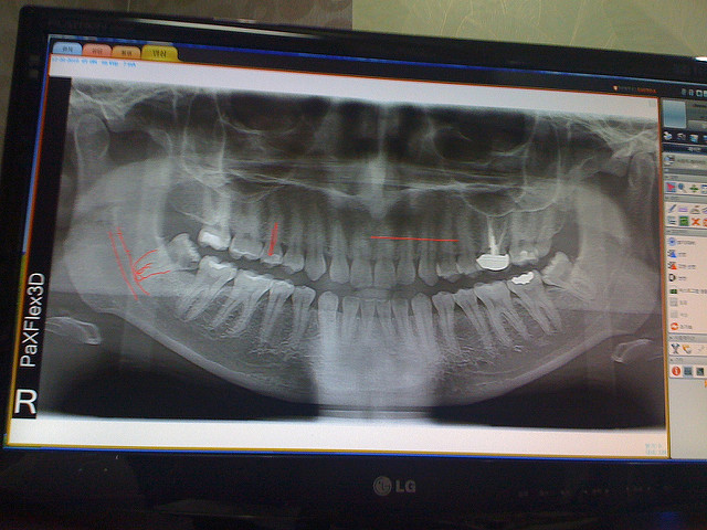
Recently, four articles that have been published on this site regarding using wisdom teeth as seen in an image from either panoramic x-ray or Magnetic Resonance Imaging (MRI) to estimate age: 1) forensic age estimation using wisdom teeth, 2) using panoramic x-rays of lower wisdom teeth to legally prove if someone is older than 18 years and 21 years, 3) Using lower wisdom teeth developmental stages determined from panoramic x-rays to calculate age, and 4) dental age estimation using MRI of wisdom teeth. From these studies discussed their appears to be a need to further explore using the so called Olze methods from Olze et al., “Evaluation of the radiographic visibility of the root pulp in the lower third molars for the purpose of forensic age estimation in living individuals,” in the International Journal of Legal Medicine, vol. 124, pp. 183–186, 2010, and Olze et al., “Assessment of the radiographic visibility of the periodontal ligament in the lower third molars for the purpose of forensic age estimation in living individuals,” International Journal of Legal Medicine, vol. 124, pp. 445–448, 2010, in conjunction with MRI and with different ethnicities. In one study previously discussed by De Tobel et al. “Forensic age estimation based on magnetic resonance imaging of third molars: converting 2D staging into 3D staging,” Annals of Human Biology, vol. 44, no. 2, pp. 121–129, 2017, the authors attempted to use the Olze method based on periodontal space (ligament) with MRI of wisdom teeth but were unable to do so in their study as they found the visibility of the periodontal ligament on MRI was insufficient to assess the stages of the Olze method in most of their images. Even so it is worthwhile to explore evidence to support the approach for use with panoramic x-ray.
In the original studies by Olze et al. of looking at both the periodontal ligament and the root pulp in a German population, the authors reached the same conclusion for both cases that if the person has reached stage 1, they must be over 18 years of age and if the person has reached stage 2, they must be over 21 years of age. In the study by Friedrich et al. also in a German population, the authors reached the conclusion for the periodontal space (ligament) that if the person has reached stage 2, they must be over 18 years of age. Thus based on both Olze et al. and Friedrich et al. one can say that if a person has reached stage 2 when assessing the periodontal space (ligament), they must be over 18 years of age. (These studies and results were previously described on this website in the earlier articles mentioned above).
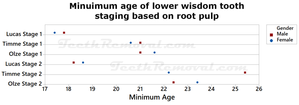
Additional studies have also been conducted to explore these methods by Olze. In a study by Timme et al. tilted “The chronology of the radiographic visibility of the periodontal ligament and the root pulp in the lower third molars,” Science and Justice, vol. 57, pp. 257–261, 2017, the authors looked at 2,346 panoramic x-rays from 1167 females and 1179 males with ages ranging from 15 to 70 years in a German population. From this initial sample, 1541 panoramic x-rays from 705 females and 836 males were used as they had sufficient quality to show at least one wisdom tooth. A total of 1,961 wisdom teeth were evaluated. Two examiners were used for the staging with the first being very experienced with dental x-rays and had received knowledge and training of assessing the visibility of the periodontal ligament from a forensic dentist and the second being a dentist without dental age estimation experience. For staging based on the periodontal ligament, the authors found the earliest onset of stage 1 was at the age of 20.1 years and 20.2 years in females and in males respectively. The authors found the earliest onset of stage 2 was at the age of 21.4 years and 26.3 years in females and males respectively for staging based on the periodontal ligament. For staging based on the root pulp, the authors found the earliest onset of stage 1 was at the age of 20.6 years and 21 years in females and in males respectively. The authors found the earliest onset of stage 2 was at the age of 22.1 years and 25.3 years in females and males respectively for staging based on the root pulp.
The authors state
“Although eruption and mineralization of third molars are the main criteria for age estimation in young living individuals, they appear not to be suitable for the validation of the completion of the 18th and the 21st year of life, even though these ages are of particular forensic importance.”
The authors in the study by Timme et al. reached a similar conclusion as the authors in Olze et al. Both males and females that had reached stage 1 were older than 18 years old, and both males and females that head reached stage 2 were older than 21 years old. Based on the results that the inter-rater agreement with the inexperienced dentist was worse than the intra-rater agreement in every case, the authors concluded that dental age estimation should only be performed by properly trained personnel. A similar conclusion was reached by De Angelis et al. “Application of age estimation methods based on teeth eruption: how easy is Olze method to use?” International Journal of Legal Medicine, vol. 128, pp. 841–844, 2014, which found considerable differences in staging results between two different physicians inexperienced with dental age estimation applying Olze’s method based on staging of the periodontal ligament to the same x-rays.
An additional study of staging the root pulp was conducted by Lucas et al. “Dental Age Estimation—Root Pulp Visibility (RPV) patterns: A reliable Mandibular Maturity Marker at the 18 year threshold,” Forensic Science International, vol. 270, pp. 98–102, 2017. The authors explored 2,000 panoramic x-rays with 1,000 females and 1,000 males in the age range from 16 to 26 years in a UK Caucasian population. The authors used a modified numbering system for staging of the root pulp visibility but the same method by Olze et al. was applied. The authors found (using the Olze et al. numbering) Stage 1 was first observed at age 17.71 years in males and at age 17.34 years in females. Stage 2 was first observed by males at 18.16 years and by females at 18.58 years. Thus the authors said that for both genders having reached stage 2 or stage 3 are unambiguous indicators that the person is over 18 years old.

A visual using the data from the root pulp staging studies of Olze et al., Lucas et al., and Timme et al. is presented below showing the minimum age of a person when the lower wisdom tooth reaches stages 1 and 2 by gender.

Thus based on the studies above it appears it can be said that for both the periodontal space (ligament) and root pulp staging methods, if a person has reached stage 2 (using numbering of the four stages from 0, 1, 2, 3) then that person must be over 18 years of age. However, these studies seem to be focused on European Caucasian populations thus caution is needed for other ethnicities. Further, the method requires a panoramic x-ray which produces a small amount of ionizing radiation. Thus additional work is needed on exploring these methods using MRI of wisdom teeth.
