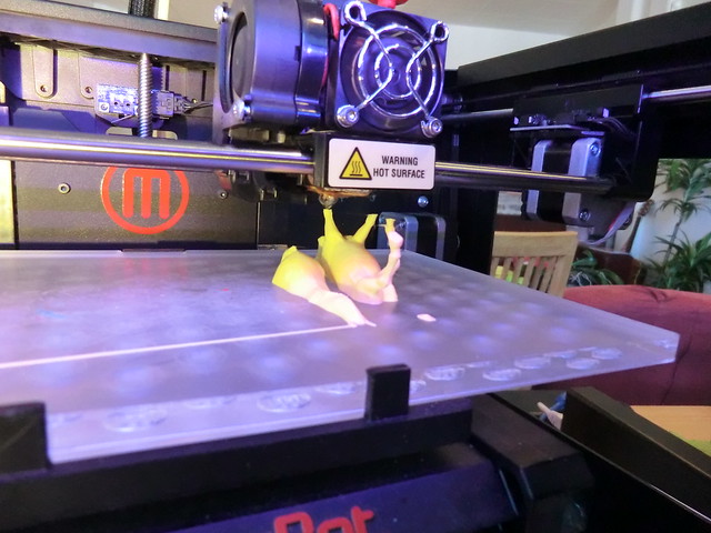
An interesting article titled “Physical Simulation Models in Oral and Maxillofacial Surgery: A New Concept in 3-Dimensional Modeling for Removal of Impacted Third Molars,” written by Cervenka et al. appears in the 2019 edition of the Journal of Oral and Maxillofacial Surgery. The article describes creating three-dimensional (3D) printed models for wisdom teeth surgery planning for particular use in residency training.
The authors discuss how advances in 3D design and printing along with decreases in costs are making the use of surgical simulation models an option for clinical training. This can have particular use with residency training because of caps on the amount of hours residents can work and increasingly patients not wanting residents to participate in their surgeries. In the article the authors present a design and fabrication of a reusable 3D printed lower jaw model with a reconfigurable wisdom tooth site that allows for as many impacted wisdom tooth configurations as one would like to explore.
In the study the authors developed a model based on a computed tomography scan of the lower jaw and used the corresponding DICOM (Digital Imaging and Communications in Medicine) file exported in 1mm slices. The authors performed thresholding on the lower jaw based on the values of the Hounsfield units and was digitally reconstructed the model using the MIMCS Innovation Suite from the company Materialise to to create a stereolithography (STL) file. The authors performed segmentation to be able to separate the wisdom teeth from the surrounding lower jaw. Eventually the authors imported the created STL file in the FreeForm Modeling Plus program produced by 3D Systems. In this software the two lower wisdom teeth were deleted and the mandible was smoothed. The lower jaw and the two wisdom teeth then had their STL files printed using software and a printer from 3D systems. It is possible to reconfigure the model in less than 1 hour. It takes 4.5 hours to print the lower jaw model and another 30 minutes to print the two wisdom teeth. The authors describe the cost of the materials as being only $50.

The authors also presents some basic information from 7 other articles that have used 3D printed models in oral and maxillofacial surgery. The authors hope that their model will help lead others in oral and maxillofacial surgery to innovate new models to use such as models of the upper jaw for the two upper wisdom teeth. The authors state
“Current applications of 3D printing in surgical training are underused; innovators have the opportunity to change the way we teach future surgeons and the way we perform surgery.”
The authors also mention how other surgical specialties have leveraged 3D printed models and particularly for educational purposes. In such cases such as with general surgery 3D models of the patient are created to allow resident to essentially rehearse the surgery in a lower pressure environment so they can be better prepared for the real surgery. Further they give an idea as to a new study that could be conducted to compare surgical times and complication rates among surgical residents who use the lower jaw and wisdom tooth model and those who do not use the model.
