An interesting article titled “Cinematic rendering to improve visualization of supplementary and ectopic teeth using CT datasets” written by Ines Willershausen and et. al. appears in Dentomaxillofacial Radiology in 2023 (no. 51, 20230058). The article discusses cinematic rendering (CR) which is a visualization technique that uses physically based volume rendering to create photorealistic images from Digital Imaging and Communications in Medicine (DICOM) data. Specifically the article attempts to tailor pre-existing CR reconstruction parameters for use in dental imaging to create 3D visualization of ectopic, impacted, and supplementary teeth.

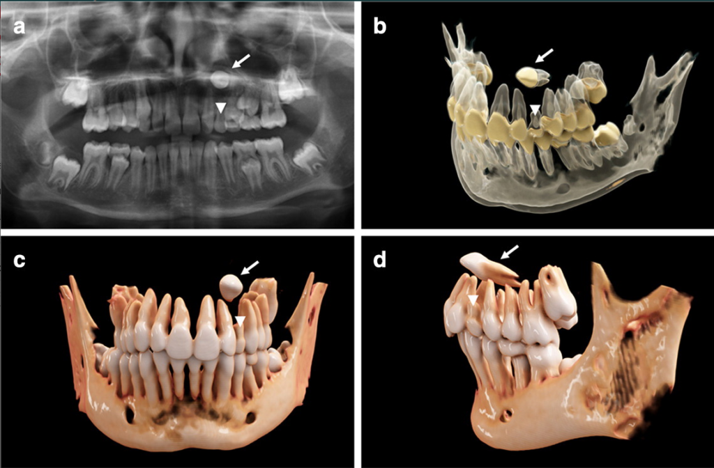
This image is from Ines Willershausen and et. al., “Cinematic rendering to improve visualization of supplementary and ectopic teeth using CT datasets,” Dentomaxillofacial Radiology, 2023, no. 51, 20230058 and has a Creative Commons license. 7-year-old girl with a horizontally impacted canine (a) A panoramic radiograph. (b) Semi-transparent reconstruction parameters are utilized to visualize bone, teeth, and dental tissues. (c, d) Frontal and lateral views of the individual dentition and bone tissue with a soft kernel.
In the article cinematic rendering was used to attempt to get a better understanding of a patient’s anatomical structures. The authors state the work performed may have been the first time this technique was used to visualize teeth. Cinematic rendering is based on data from CT and cone-beam CT images, and the visualization technique uses physically based volume rendering to create photorealistic images from DICOM images. Cinematic rendering builds upon physically based light simulation algorithms in which billions of virtual light rays are simulated every second. The end result is an appearance with images with more realistic than conventional volume rendering.

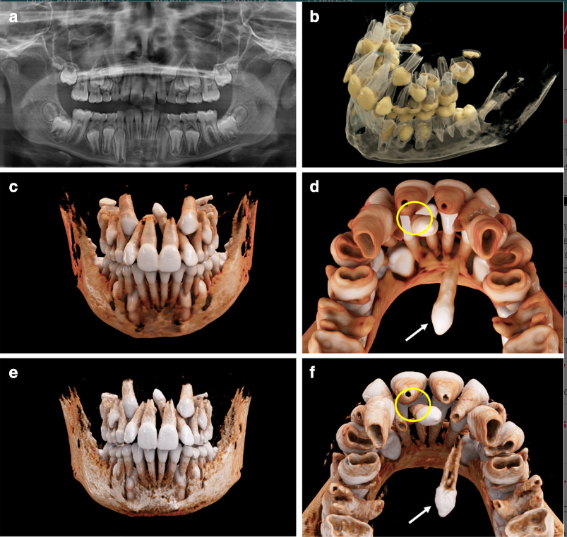
This image is from Ines Willershausen and et. al., “Cinematic rendering to improve visualization of supplementary and ectopic teeth using CT datasets,” Dentomaxillofacial Radiology, 2023, no. 51, 20230058 and has a Creative Commons license. 11.2-year-old boy with a mesiodens and a further supplementary tooth (a) A panoramic radiograph. (b) Semi-transparent reconstruction parameters are utilized to visualize bone, teeth, and dental tissues. (c, d) Views of the individual dentition and bone tissue with a soft kernel. (d, e) Views of the individual dentition and bone tissue with a hard kernel.
So far there has been limited research on this cinematic rendering in dentistry. At the time of publication of the article, cinematic rendering has focused on fractures and carcinoma as maxillofacial indications. In the article the researchers used cinematic rendering for the volumetric image visualization from CT datasets of the midface. Predefined reconstruction parameters were modified to visualize complex orthodontic cases in kids with ectopic, impacted, and supernumerary teeth. The researchers used the masking and windowing features of Siemens Healthineers’ Cinematic Anatomy application. This allowed them to create segmentations of the upper and lower jaw and respective dentitions in order to create natural-appearing images for realistic representations of anatomy. The authors noted that, the reconstruction time can take up to 24 hours and thus is not yet ready for routine clinical use. Reconstruction time is expected to significantly decrease over time as the software improves and better hardware is used when running the software. Even so the authors feel that the 3D spatial relationship of the teeth, and structural relationship with the antagonizing dentition, can be shown as an interactive 3D visualization after segmentation through windowing. The authors state:
“To the best of our knowledge, CR has not been implemented for the visualization of supplementary and ectopic teeth segmented from the surrounding bone because the software has not yet provided appropriate customized reconstruction parameters for dental imaging. When employing our new, modified reconstruction parameters, its application presents a fast approach to obtain realistic visualizations of both dental and osseous structures.”

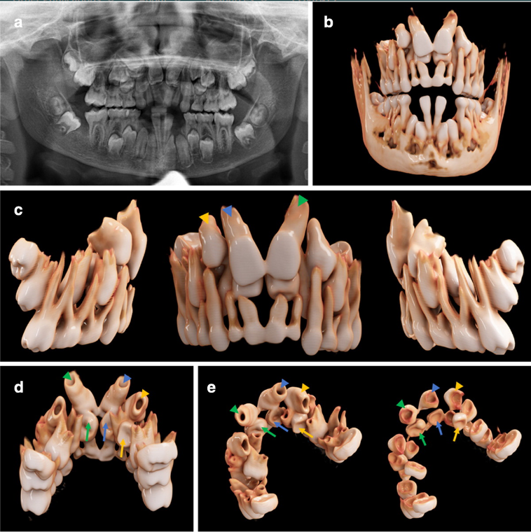
This image is from Ines Willershausen and et. al., “Cinematic rendering to improve visualization of supplementary and ectopic teeth using CT datasets,” Dentomaxillofacial Radiology, 2023, no. 51, 20230058 and has a Creative Commons license. 12.4-year-old boy with cleidocranial dysostosis and multiple supernumerary and impacted teeth. (a) A panoramic radiograph. (b-e) The bone present with a soft kernel in the frontal, lateral, and palatal view.
The authors feel that CR will enabe dentists and oral surgeons to gain an improved 3D understanding of anatomical structures, and this can enable more intuitive treatment planning and patient communication.
