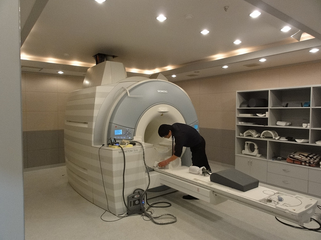
Recently, three articles have been published on this site regarding using wisdom teeth to estimate age: 1) forensic age estimation using wisdom teeth, 2) using panoramic x-rays of lower wisdom teeth to legally prove if someone is older than 18 years and 21 years , and 3) Using lower wisdom teeth developmental stages determined from panoramic x-rays to calculate age. All such articles use panoramic x-rays of wisdom teeth in order to attempt to estimate the age of the person they came from. However, recently there has also been articles describing using Magnetic Resonance Imaging (MRI) of wisdom teeth to predict age.
One article is by Baumann et al. “Dental age estimation of living persons: Comparison of MRI with OPG,” Forensic Science International, vol. 253, pp. 76–80, 2015. Another article is by Guo et al. “Dental age estimation in living individuals using 3.0 T MRI of lower third molars,” International Journal of Legal Medicine, vol. 129, pp. 1265–1270, 2015. Yet another article is by De Tobel et al. “Forensic age estimation based on magnetic resonance imaging of third molars: converting 2D staging into 3D staging,” Annals of Human Biology, vol. 44, no. 2, pp. 121–129, 2017.
In the article by Baumann et al. the motivation was due to migration of adolescents from countries where birth dates are not reliably documented. An additional motivation is because of ionizing radiation by panoramic X-ray which is ethically controversial. MRI is not based on X-rays and does not cause any ionizing radiation. Dental MRI is not often used in dentistry. The researchers set out to determine if the assessment of the mineralization and the eruption of the wisdom teeth used with panoramic x-ray can be applied to MRI. In the study twenty-seven (27) caucasians with 19 females and 8 males living in Austria with a known age between 13 and 24 years with at least two present wisdom teeth had an MRI scan of the jaw performed within 14 days after a panoramic x-ray. The MRI was performed using a 3 Tesla (T) scanner with an acquisition time of just under 21 minutes. Two dentists independently read the MRI and panoramic x-rays scans. Mineralization and eruption stages of the molars of all for quadrants were evaluated. Mineralization was assessed according to the staging system established by Demirjian et al. and eruption was evaluated according to the staging by Olze et al. (described more in the previous articles on this website).
In the article by Baumann et al. 262 molars were assessed for the mineralization
and 274 for the evaluation of eruption, in both panoramic x-ray and MRI data. The dataset initially had 312 molars but 5 were missing and some other molars were not assessed due to imaging artifacts. The authors found that MRI can be used for dental age estimation about as well as panoramic x-ray. Further, both, mineralization and eruption stages were able to be identified equally well in both imaging modalities. For mineralization the authors found that MRI allowed a better evaluation of the stages which the authors speculated could be because the roots of the molars could be superimposed in panoramic x-ray. For eruption the authors found that panoramic x-ray allowed a better evaluation of the stages which the authors felt could be be due to the evaluation of eruption involving an assessment of the dental crown in the oral cavity which is prone to metallic artifacts from dental retainers in MRI. The researchers also found for both, mineralization and eruption, there was a tendency of assessing slightly higher stages in the panoramic x-ray. Overall the authors felt MRI of wisdom teeth can be used for age estimation instead of using panoramic x-rays.
In the article by Gao et al. the authors studied 3.0 T MRI scans taking just under 6 minutes long of wisdom teeth from 517 Germans with 269 males and 248 females aged 12 to 24 years. The authors studied mineralization stages of lower wisdom teeth according to staging by Demirjian et al. The authors started with 613 Germans but reduced this amount to 517 because some had no lower wisdom teeth and in some images the mineralization could not be reliably assessed. The authors were interested in the lower left wisdom tooth but in cases where the lower left wisdom tooth had been extracted the authors looked at the lower right wisdom tooth. The authors compared means and standard deviations in terms of years in males and females individually for mineralization stages B to H of lower left wisdom teeth to two prior studies that used panoramic x-rays. Slight differences between studies were found which was explained by differences in the age ranges and age distributions used and also the factor that different imaging modalities were used. The authors found that in stages C, E, F, and G there was a statistically significant lower mean ages in males than in females. Thus the authors found a faster developmental rate for males in several mineralization stages which the authors noted is in agreement with prior studies. From their results the authors feel that MRI is an alternative to panoramic x-ray in assessing wisdom teeth mineralization.
In the article by De Tobel et al. the authors studied 3.0 T MRI scans of 52 Belgians aged 14 to 26 years with 16 of them also having had a panoramic x-ray taken within 6 months that was available. All images were assessed by two observers that were trained in dental age estimation. The wisdom teeth were staged accordingly to methods by Demirjian et al. An attempt was also made to stage using the method described by Olze et al. (as discussed in using panoramic x-rays of lower wisdom teeth to legally prove if someone is older than 18 years and 21 years) but the periodontal ligament was not able to be assessed with MR. In some cases an evaluation of a root was impossible on the panoramic x-ray due to superposition of other anatomical structures. In some cases on there insufficient image quality or contrast on MR to assess the root. The authors found the number of assessable upper wisdom teeth on MR (62 out of 64 – 97%) was statistically significantly different from the number on panoramic x-ray (37 out of 64 – 58%). However there was not a similar statistically significantly difference for lower wisdom teeth. The authors also found that for when a upper wisdom tooth appeared assessable the staging was statistically significantly less difficult when based on MRI. However, no similar statistically significant difference was found when comparing lower wisdom teeth with MRI and panoramic x-ray. The authors also found that no matter which imaging technique was used, the staging of the lower wisdom teeth was associated with less variability than the staging of the upper wisdom teeth. The authors also found a better agreement between the two observers on MRI than on panoramic x-ray. The authors also found that the palatal root was most suitable for staging in upper wisdom teeth and the distal rot was most suitable for staging in lower wisdom teeth.
In the article by De Tobel et al. the authors note four limitations of their study. The first is due to a small sample size used of panoramic x-rays that could be compared to MRI. Another limitation was that when both panoramic x-rays and MRI were available that was often much time taken between the images that ideally should be taken on the same day. Another limitation was the 2mm slice thickness of MRI used which could cause insufficient resolution. A final limitation was there was no investigation of the influence of impaction and of extracted premolars performed. Nonetheless, the authors feel that using MRI of wisdom teeth can be used for age estimation.

In all three articles Baumann et al., Guo et al., and De Tobel et al. the authors feel that MRI of wisdom teeth can be used for age estimation instead of using panoramic x-ray. Even so it appears that this would probably be more costly and have a greater scan time. In addition, the staging approaches used for panoramic x-ray may need to be refined for use with MRI. It also appears from the work by De Tobel et al. that MRI can be better used for staging the upper wisdom teeth which has often been omitted in prior studies. This might allow for more accurate predictions of age by considering all the wisdom teeth and not just the lower wisdom teeth.

1 thought on “Dental Age Estimation Using MRI of Wisdom Teeth”