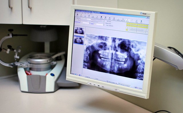
An interesting article titled “Is Panoramic Imaging Equivalent to Cone-Beam Computed Tomography for Classifying Impacted Lower Third Molars?” appears in the 2019 edition of the Journal of Oral and Maxillofacial Surgery written by Brasil et al. The article explorers if panoramic radiography is able to give similar results as cone-beam computed tomography (CBCT) for the degree of lower wisdom teeth impaction and when using panoramic radiography if the external oblique ridge is a reliable indicator for the degree of lower wisdom teeth impaction.
In the article the authors discuss the Pell and Gregory classification which is used to classify the position of a wisdom tooth and can be used to potentially assess the risks of various complications. The authors state how wisdom teeth are usually evaluated using panoramic x-rays but this technique is susceptible to image overlap, magnification, and distortion, and particularly in the ascending ramus of the mandible which is needed for proper classification using the Pell and Gregory classification. As an alternative cone-beam computed tomography (CBCT) images can provide 3D views of the wisdom tooth and surrounding regions.

The authors devised a study using 173 patients with 313 lower wisdom teeth. The patients were required to have their at least one lower wisdom tooth imaged using both panoramic radiograph and CBCT within 15 days of each other. ImageJ software was used by two oral and maxillofacial radiologists to classify the wisdom teeth using the panoramic radiographs. OnDemand3D software was used by the two oral and maxillofacial radiologists to classify the wisdom teeth using CBCT. Thirty days after performing the evaluations, 20% of the sample of images was selected and re-evaluated in order to verify that similar results as before were obtained. The authors found an overall agreement rate of 87.26% between the two imaging modalities. Further, there was agreement between the two imaging modalities for Pell and Gregory class A, B, and C of 82.1%, 90.5%, and 65.6% respectively. The authors found that the anatomical landmarks for a reference for panoramic radiographs was statistically significantly different than that for CBCT with an agreement rate ranging from 66.8% to 76.4%. The authors found that when such disagreement occurred the space to accommodate a wisdom tooth was found to be underestimated in panoramic radiographs.
The typical reference landmark used for the Pell and Gregory classification of wisdom teeth is the ascending ramus of the mandible. The authors also explored using the external oblique line as the reference landmark but it was found to underestimate the space to accommodate wisdom teeth for panoramic radiographs and not be reliable as a landmark. The authors state
“For both landmarks on PR [panoramic radiograph], such underestimation of the available space (or overestimation of the impaction degree) could mislead the professional, causing him or her to overestimate the level of difficulty of the surgical procedure.”
The authors state that even though images provided by CBCT can be more reliable for evaluating tooth impaction it should not be routinely used to plan wisdom teeth surgery. This is because in many cases CBCT may not yield any improved diagnosis capability and thus the patient would receive more ionizing radiation for no benefit. However, in some cases the use of CBCT is justified prior to surgical removal of wisdom teeth. The authors also state:
“According to the results of our study, mandibular ramus impaction using PR might not represent the true relative position of the tooth and its related complication risks; therefore, those must be cautiously interpreted.”
Thus the authors feel that additional studies should be conducted using CBCT to classify wisdom teeth impaction and assess the risk of complications such as mandibular angle fractures and cavity development on the adjacent second molars. This will allow to see if panoramic radiograph can be considered reliable for predicting such complication risks of wisdom teeth removal.
