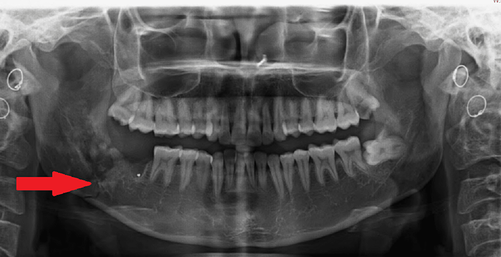
An interesting article titled “Iatrogenic Fracture of the Mandibular Angle During Excision of an Impacted Third Molar” appears in Cureus in August 04, 2022, written by Agrawal P, Jadhav A, Bhola N D (vol. 14, no. 8, pp. e27672). The article discusses a case of a 30 year old woman who had atrogenic mandibular angle fracture after having an impacted wisdom tooth removed.
In the article, the authors mention of a 30 year old woman visited their outpatient department complaining of pain and swelling over the lower right side of her face with an inability to open her mouth and chew for the last four days. Prior to this, she had her lower right impacted wisdom tooth removed under local anesthesia. On exam, the woman was found to have an asymmetrical face due to swelling over the lower right region of the face and her skin was tender on palpation. An panoramic x-ray was taken which showed a thin radiolucent line running obliquely from the alveolar crest of the extraction site to the angle of the mandible, indicating a fracture. As a result, a computed tomography (CT) scan was also taken which showed a tooth socket with gross destruction of the lingual cortical plate and a breach in the continuity of bone through the socket, which suggested a mandibular angle fracture.
The woman had her swelling treated and then the fracture was treated with open reduction and two-point fixation using a trans-buccal approach with titanium plates and screws. She was tehn give antibiotics and pain killers and discharged after five days. The woman recovered without any reported issues. At a six month follow up the woman had a functionally stable occlusion.
In the discussion of the case, the authors mention how mandibular fracture a known issue of wisom teeth removal but very rare and is believed to occur in between 0.0034% and 0.0075% of all wisom teeth surgeries. A mandibular angle fracture can occur as it is one of the vulnerable regions prone to fracture due to complex biomechanics and anatomical characteristics. An impacted wisdom tooth is beleived to decrease the cross-sectional area of bone and leads to a two to four time increase in fractures of the mandibular angle. These types of fractures are difficult to treat because the masseter and medial pterygoid muscles are attached to the angle. Further, pain, swelling, and restricted mouth opening can make examantion challenging. It is beleived that age, gender, bone present, the position of the tooth, the volume of the tooth, the depth of impaction, and systemic conditions can influence bone metabolism which can contribute to a mandibular angle fracture. A CT scan is considered the gold standard to detect a mandibular angle fracture which was performed in this case after an initial panoramic x-ray.

Jaw fractures can occur from excessive force and incorrect use of instrumentation such as elevators or forceps during surgery. The authors also mention that IFM could occur due to the presence of former bony pathologies such as cysts and tumors, recurrent pericoronitis, and periodontal disease which can weaken the mandible and lead to a predispotion towars fracture. It is also note that surgeons with less experience are also more likely to cause such a fracture. When a mandibular angle fracture occurs, patients have different treatment options including intermaxillary fixation to open reduction and internal fixation, a soft diet, and even no treatment.
The management strategies for mandibular angle fractures are diverse and range from no treatment, soft diet, and intermaxillary fixation to open reduction and internal fixation. Treatment options are dictated by the characteristics of a fracture and the surgeon’s preference. In the present case, after explaining all the treatment options, the patient opted for open reduction over intermaxillary fixation. The authors state:
“A detailed preoperative evaluation of the impacted teeth and bone adjacent to the sectioning of the tooth and minimal removal of bone during trans-alveolar extraction are a few techniques that can be employed to avoid [mandibular angle fracture].”
The authors therefore suggest that surgeons should well aware of patient anatomy and how to use elevators and forceps when sectioning a tooth during wisom teeth surgery to help avoid mandibular angle fracture. It seems as if additional studies are needed on mandibular angle fracture after wisdom teeth surgery to provide more evidence for or against potential contributing factors of anklosis of tooh, age, gender, reduction of the elasticity of bone, narrowing of the periodontal ligament space, and greater masticatory force.
