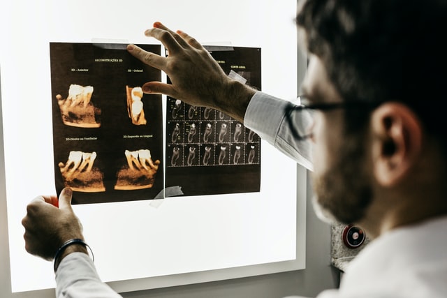
An interesting study titled “Does clinical experience with dental traumatology impact 2D and 3D radiodiagnostic performance in paediatric dentists? An exploratory study” appears in the BMC Oral Health written by Gertrude Van Gorp and et al. (vol. 22, no. 245, 2022). The article seeks to explore the performance of pediatric dentists when identifying and detecting traumatic dental injuries on both 2D and 3D images.
In the study the authors analyzed data from nine pediatric dentists (six female and three male) who had used structured scoring sheets to randomly assess 2D and 3D images of anterior permanent teeth with dental trauma. The researchers analyzed the level of experience with traumatic dental injuries on imaging, identification, and interpretation of lesions. The authors compared these results to benchmark data from expert consensus of an experienced dentomaxillofacial radiologist and pediatric endodontist. Six of of the pediatric dentists in the study were classified as having low traumatic dental injury experience while the other three had a high level of experience. Further, most dentists in the study had no experience with cone-beam computed tomography (CBCT). CBCT is often used when more severe injuries occur as it can allow for a more precise diagnosis but is more difficult to interpret than x-rays. Six of the dentists included in the study had no formal training on CBCT.
The authors found that the performance of the pediatric dentists was moderate to poor and sensitivity percentages on detection, identification, and image interpretation or both 2D and 3D images were in the range of 34.8% to 56%. Only around half of the radiographic findings recorded by the benchmark data were detected by the pediatric dentists. Further less than half of the radiographic findings were identified correctly. The authors feel that this indicates that the assessment of the radiographic images was incomplete and suboptimal and there was possible risk of overlooking items relevant for a correct diagnosis and identification of the appropriate treatment plan. In addition the authors found there was no statistically signficant diference found between those pediatric dentists with low and high traumatic dental injury expeirence for the three outcomes measures and two types of imaging types. Also, the authors found that 3D imaging improved detection and identification of findings in those dentists with low traumatic dental injury experience but feel that 3D imaging cannot compensate for the lack of clinical experience. In general the authors feel that pediatric dentists need more CBCT-related training to to cover potetial knowledge gaps.
The authors do note that the study was limited by the sample size and thus a lack of statistical power and only having nine pediatric dentists in the analysis. Another study limiation was that the authors retrieved findings from patient records and then periapical radiographs were presented to the dentists in a power point presentation and not in a true clinical setting, although this was not the case for the CBCT data.

As such, the authors caution that the results should be considered exploratory. Even so, the authors state:
“The impact of this research fnding needs further exploration, but it is clear that paediatric dentists could beneft from more training in the assessment and interpretation of radiographic images, both 2D and 3D.”
The authors feel further studies would be beneficial and suggest a follow up study to include what treatment the dentists would propose for each trauma case,
based on the 2D and 3D images and to expore the possible impact on such a treatment. In their conclusion, the authors even go as far as to suggest that image interpretation in a pediatric dentistry setting may be best left to an experienced dento-maxillofacial radiologist.
