Recently on this website research exploring using photoacoustic imaging for periodontal probe depths from University of California, San Diego, using swine models was discussed. This same group has since published an article titled “Photoacoustic imaging for monitoring periodontal health: A first human study,” by Moore et al. in Photoacoustics (vol. 12, pp. 67-74, 2018, published online November 01, 2018) where they show that a photoacoustic-ultrasound imaging approach can image the full depths and geometries of pockets in healthy human adults. Traditionally ultrasound uses the principle sound in and sound out but the photoacoustic-ultrasound approach uses light in, sound out. The advantageous of such an approach over traditional radiography is that it can image soft issue and that it does not cause ionizing radiation.
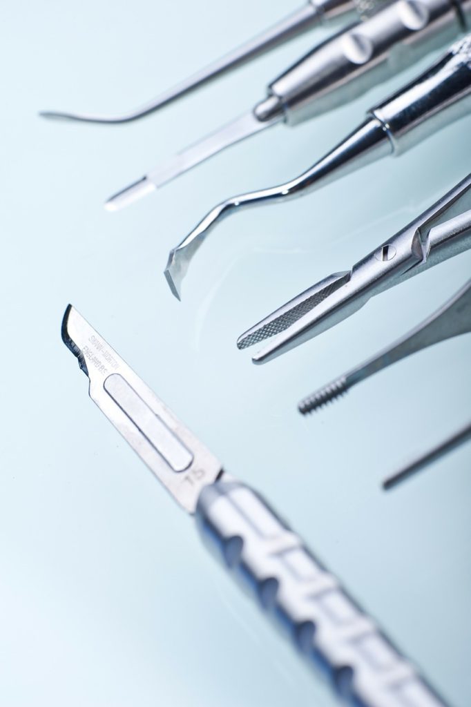
The conventional method for dentists to monitor gingival health in humans is with a periodontal probe. This instrument is a thin, hook-like metal tool that is marked like a tiny measuring stick and inserted in between the teeth and gums to see if the gums have shrunk back from the teeth, creating pockets. The researchers stated that the periodontal probe is a relatively unsophisticated tool that has poor reproducibility means measurements vary greatly between dentists. Furthermore, the probe is only capable of measuring the pocket depth at the point of insertion. It is also possible that the probe can led to patient discomfort and bleeding by penetrating inflamed tissue. Being able to assess the periodontal probe depth is said to be important for assessing wisdom teeth in patients. A periodontal probing depth of greater than or equal to 4 mm is used by the American Association of Oral and Maxillofacial Surgeons (AAOMS) as a cutoff for when they feel wisdom teeth should be extracted due to the increased risk of potentially developing periodontal disease. In general a pocket depth measuring up to two millimeters (mm) indicates healthy gums while a pocket depth measuring three mm and up is an indicator of gum disease.
For the current study, the researchers enrolled a healthy 22 year old female with good oral hygiene. First a convention periodontal probe was used to measure pocket depths. All measurements less than 2 mm were recorded as 2 mm. Next a contrast agent was prepared from cuttlefish ink and the subgingival pockets were labeled. Photoacoustic imaging was performed by pulsing light through two optical fiber bundles. Three transducers then collected the sound data. Next both a photoacoustic and ultrasound image were analyzed to determine where both images were above a 4% minimum pixel intensity to then consider this point as part of the pocket depth.
The researchers compared the measurements taken from the periodontal probe and their photoacoustic imaging approach. For teeth numbers 24 and 25, the pocket depth was rounded up to 2 mm from measurements taken from the
periodontal probe. For these same two teeth with the imaging approach pocket depths were found to be 1.34 mm and 1.15 mm. The researchers also took several experiments to demonstrate reproducibility by performing their described labelling, imaging, cleaning, and processing procedure to assess pocket depths on different days. The researchers found that the mean standard deviation of their measurements taken to assess the full geometry of pockets was 10.2%.

The researchers state that their approach offers a major improvement over the standard periodontal probe and that it can be accurate to reveal the buccal pocket with sub-millimeter precision. One limitation with their study was that the geometry of the transducer physically limited access to all 32 teeth and so further work is needed for a custom mouthpiece shaped device. In the study only teeth numbers 7 to 10 and 22 to 27 were assessed. It is also important to note that the woman that was imaged did not have any deep pockets greater than 4 mm. Wisdom teeth have teeth numbers 1, 16, 17, and 32 and therefore were not assessed in this study. Thus further work is needed to be done to better assess if this approach can be used for helping assess if wisdom teeth should be extracted or retained.
