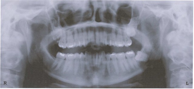
An interesting article titled “Predictive Value of Panoramic Radiography for Injury of Inferior Alveolar Nerve After Mandibular Third Molar Surgery,” appears in the 2017 edition of the Journal of Oral and Maxillofacial Surgery (vol. 75, pp. 663-679) written by Su et al. The article sought to explore if panoramic x-rays taken before wisdom teeth removal can predict possible injury of the inferior alveolar nerve.
In the article the authors discuss how inferior alveolar nerve injury is the third most common complication following wisdom teeth removal. When inferior alveolar nerve injury occurs numbness of the lip or chin can occur along with difficulty speaking and chewing food. Current studies have shown inferior alveolar nerve injury occurs about 8% of the time after wisdom teeth removal with less than 1% expected to be permanent. Current practice has shown that panoramic x-rays can provide visualization of the proximity of inferior alveolar nerve canal and molar roots to wisdom teeth. Seven radiographic signs have been suggested in the past for prediction of inferior alveolar nerve injury after wisdom teeth removal including: 1) darkening of the root, 2) deflection of the root, 3) narrowing of the root, 4) dark and bifid apex of the root, 5) diversion of the canal, 6) interruption of white line of the canal, and 7) narrowing of the canal. The authors devised a way to systematically review the diagnostic predictive accuracy of these 7 panoramic radiographic signs for the risk of inferior alveolar nerve injury after wisdom teeth removal as they stated a comprehensive review had not yet been done.
The authors conducted a search of studies related to using at least one of these 7 radiographic signs with elective wisdom teeth removal to predict inferior alveolar nerve injury. The authors also only included studies where false-positive, true-positive, false-negative, and true-negative proportions for the occurrence of
inferior alveolar nerve injury after wisdom teeth removal were recorded for the radiographic signs or the authors were able to calculate these from the data. This allowed the authors to re-calculate the positive predictive value (risk of inferior alveolar nerve injury after wisdom teeth removal with positive radiographic signs), negative predictive value (risk of absence of inferior alveolar nerve injury after wisdom teeth removal with positive radiographic signs), sensitivity (the risk of
inferior alveolar nerve injury after wisdom teeth removal was identified correctly using radiographic signs), and specificity (the risk of absence of inferior alveolar nerve injury after wisdom teeth removal was identified correctly using radiographic signs).

Eventually the authors arrived at 8 studies meeting their inclusion criteria for meta-analysis. Between 4 of these 8 studies and all 8 of the studies were pooled for each of the 7 radiographic signs to calculate the pooled prevalence of experiencing an
inferior alveolar nerve injury along with the positive predictive value, negative predictive value, sensitivity, and specificity. Specifically the prevalence of experiencing inferior alveolar nerve injury after wisdom teeth removal based on diversion of canal was 4.4%, based on interruption of white line of the canal was 4.8%, based on narrowing of the canal was 4.2%, based on darkening of the root was 4.5%, based on deflection of the root was 4.1%, based on narrowing of the root was 4.2%, and based on dark and bifid apex of the root was 4.1%. The study showed the negative predictive values for all 7 radiological signs were too low for ruling out the risk of inferior alveolar nerve injury prior to having wisdom teeth removed. However the authors found the positive predictive values for the presence of diversion of the canal, interruption of the white line of the canal, and darkening of the root to be able to show a risk inferior alveolar nerve injury after wisdom teeth removal.
The authors state
“For an OMS [oral and maxillofacial surgeon], if a patient has a positive panoramic radiographic screening result for 1 of these 3 signs, the patient will have an 8 to 22% greater risk of experiencing a postoperative IAN [inferior alveolar nerve] injury. Hence, an OMS
[oral and maxillofacial surgeon] could consider a more conservative method such as coronectomy for the management of mandibular MM3 [lower wisdom teeth] to prevent an IAN [inferior alveolar nerve] injury or further pursue the 3-dimensional relationship between the MM3 [ wisdom teeth] root and the IAN [inferior alveolar nerve] before the procedure.”
The authors found that for the other 4 radiographic signs, specifically, deflection of the root, narrowing of the root, dark and bifid apex of the root, and narrowing of the canal, the positive predictive values and negative predictive values were very close or even less than the prior probabilities which indicated these signs were very small or even absent and may not predict accurately a possible inferior alveolar nerve injury after wisdom teeth surgery. The authors did point out several issues such as when they did their calculations of positive predictive values and negative predictive values they treated each of the 7 radiographic signs as independent in two of the eight studies used. In addition some of the eight studies included in their analysis were described as having major design flaws by the authors. Ultimately the study can help provide some information to patients regarding their potential risk of inferior alveolar nerve injury if they were to have wisdom teeth removal. Additional higher quality studies are needed to better assess the 7 radiographic signs.
