An interesting article titled “Marsupialization of Dentigerous Cysts Followed by Enucleation and Extraction of Deeply Impacted Third Molars: A Report of Two Cases” appears in Cureus in April 02, 2022 written by Nedal Abu-Mostafa (vol. 14, no. 4, pp. e23772). The article discusses two cases of a dentigerous cyst (DC) that involves the crown or a portion of the crown of an unerupted or impacted tooth that more commonly affect mandibular or lower impacted wisdom teeth.
In the article discussion is made of dentigerous cysts being the second most common odontogenic cysts. Dentigerous cysts are caused by an alteration of the reduced enamel epithelium and results in fluid accumulation between it and the enamel of the crown. The progression of dentigerous cystsis are slow and often do not present any symptoms and the patient learns about them through an x-ray or looking into reasons why a tooth failed to erupt or had misalignment. Treatment can occur through enucleation or marsupialization. Enucleation is a surgery that extracts the affected tooth and shelling out the entire cystic lining. Marsupialization is when a window is formed on the cystic wall, the cyst fluid is evacuated then the cystic lining is sutured to the oral mucosa. Marsupialization is therapy for large cysts that are close to structures like the maxillary sinus and inferior alveolar nerve. Marsupialization may not be able to cure the cystic cavity completely or stimulate the cyst-associated tooth to fully erupt and may need to be followed by enucleation. In the article discussion is made of two cases of deeply impacted wisdom teeth with dentigerous cysts that were treated with marsupialization followed by an atraumatic surgical extraction and enucleation of the residual lining.
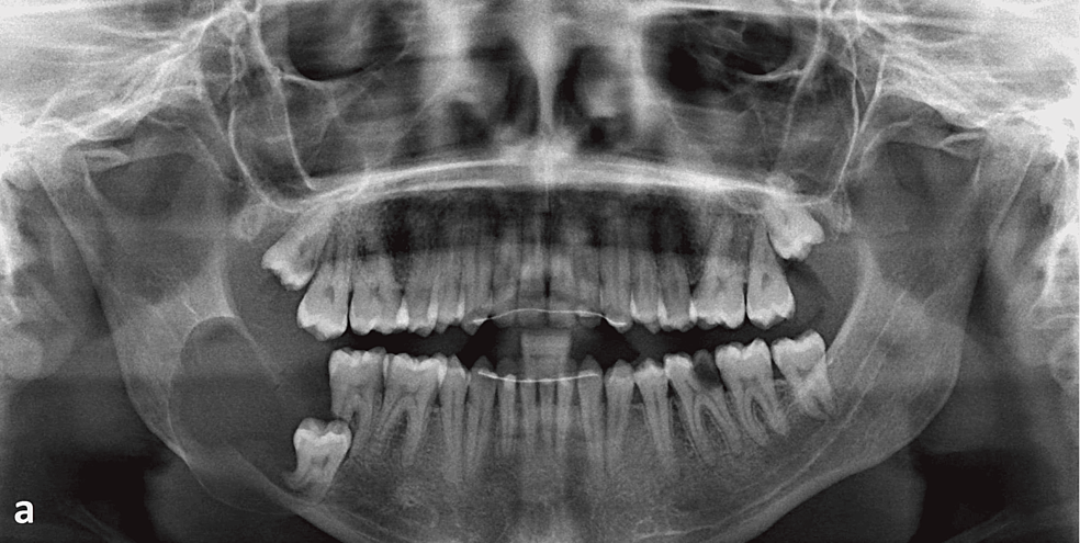
In the first case in July 2018, a 25-year male patient complained of swelling on the right side of his lower jaw that had started three months prior and had grown in size. A panoramic radiograph showed a large unilocular radiolucent lesion enveloping the crown, distal and mesial roots of the lower right wisdom tooth. The steps of marsupialization were explained to the man including creating a window in the cystic cavity, removal of a portion of the lining for biopsy, and packing the cavity with gauze. The man consented to the procedure but did not come to his next scheduled appointment and returned six months later. The procedure was then performed under local anesthesia and two weeks after surgery the wound healing was found to be adequate. The patient came into the office every 3 days for two weeks to have his gauze pack changed before being given a syringe to irrigate the cavity twice daily with regular saline. After 8 months a new panoramoic x-ray showed a reduction in the distal and inferior borders of the cystic cavity with a limited occlusal and distal movement of the wisdom tooth and surgical extraction was performed. Fifteen months after the extraction, the man had complete bone healing at the extraction site and the man’s lower lip and skin had normal sensations.

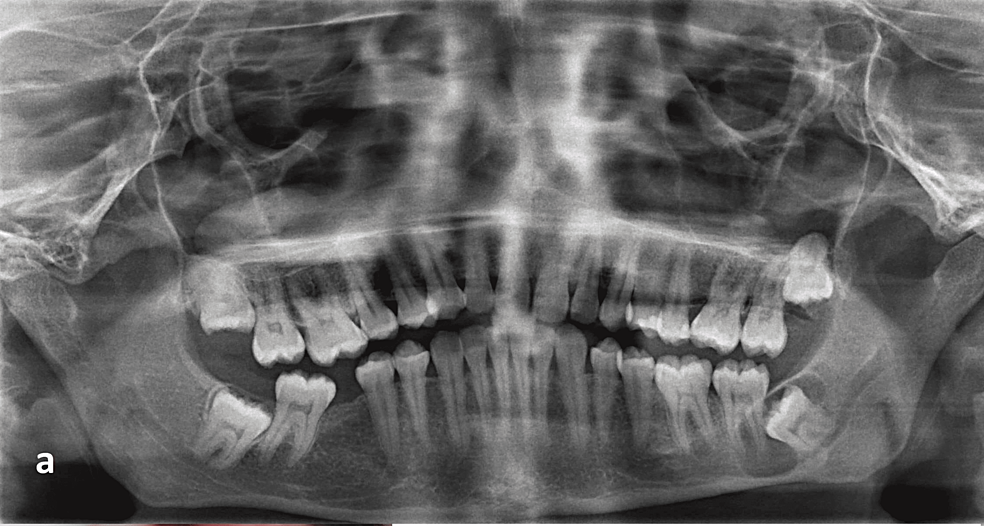
In the second case in January 2019, a 36-year old male patient came in for dental care and had a panoramic radiograph taken which showed a mesioangular impacted lower left wisdom tooth with divergent roots near the inferior alveolar canal along with a radiolucent lesion with a radio-opaque border which suggested a dentigerous cyst. Enucleation was ruled out as a treatment option and marsupialization was suggested to shift the cyst lesion followed by later extraction. The procedure was accomplished using similar techinque as that in the first case with complete details described in the article. A panoramic radiograph was taken four months after marsupialization which showed the cyst had shrunk but surgical extraction was postponed. After eight months another panoramic radiograph was taken which showed a small radiolucency anterior to the mesial wall of the wisdom tooth crown and posterior to the distal root of the second molar and enucleation and surgical extraction was performed. The man’s lower lip and skin had a normal sensation and after 14 months the man had complete bone healing at the extraction site.

When discussing the cases the author states:
“Treatment of DCs associated with third molars [wisdom teeth] varies from similar cysts that involve other teeth, as saving third molars in the dental arch is not necessary….The recommended therapy is marsupialization, with the goal of moving the cystic cavity and the implicated third molar away from the canal before attempting to remove the cyst lining. The tooth in concern may erupt on its own, be extruded by orthodontic treatment, or be surgically removed.”
The two cases described had an initial treatment of the cysts with marsupialization followed by extrusion of the cyst-associated teeth and then finally extraction.
