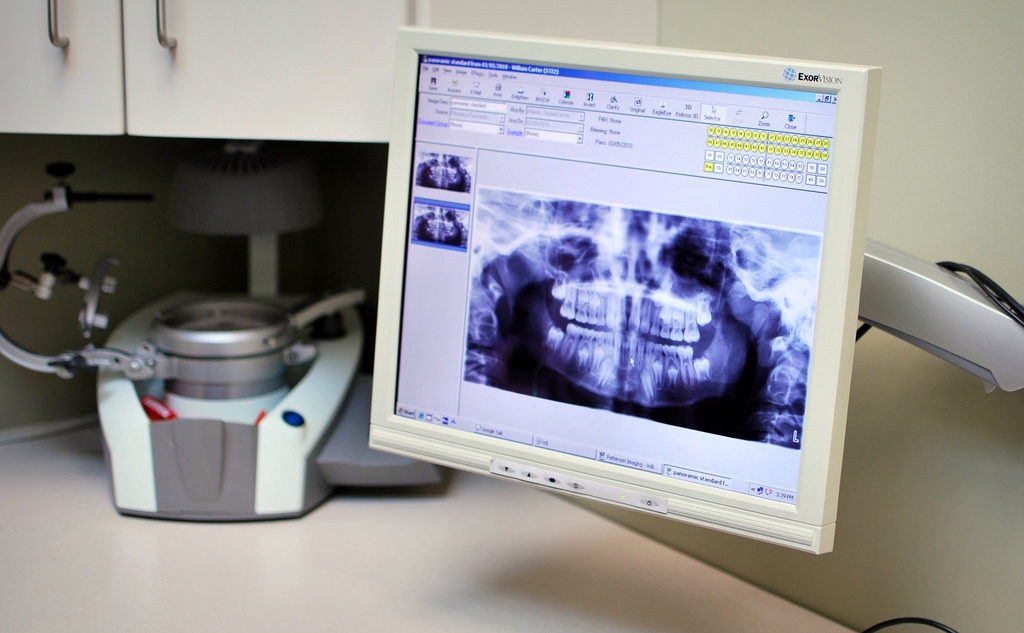
An interesting article titled “Impaction of lower third molars and their association with age: radiological perspectives,” written by Ryalat et al. appears in BMC Oral Health in 2018 (vol. 18, no. 1, Published April 4, 2018). The article explores the impaction pattern in radiographic images of wisdom teeth. The authors were motivated by the belief that if the impaction patterns of wisdom teeth can be identified this can help guide clinical decision making regarding to extract or to retain lower impacted wisdom teeth.
Specifically, for the study two individuals looked at 1,198 orthopantomographs or
panoramic radiographs with 1,810 impacted lower wisdom teeth or third molars. Originally a total of 4,600 orthopantomographs were retrieved from those taken at The University of Jordan Hospital between the years 2010 and 2014, but 3,402 were excluded due to patient age being outside of the range of 18 to 26, patients with craniofacial anomalies, patients who were undergoing orthodontic treatment such as braces, fully erupted wisdom teeth, and a few other reasons as described in the article. The researchers selected the age range of 18 to 26 based on prior research that shows this group is the most likely to show significant movement in lower wisdom teeth.
The researchers defined a wisdom tooth’s angulation as the angle formed between the dental long axis and occlusal plane where horizontal is defined as less than 20°, mesioangular is defined as between and including 20° and 80°, vertical
is defined as between and including 80° and 100°, and distoangular is defined as more than 100°. The study used software from Kodak which made the machine taking the images to help calculate angles. They also used SPSS for statistical analysis.
The results showed that the majority of impactions were mesioangular (66.1%) followed by vertical impactions (18.8%) and then horizontal impactions (15.1%). The radiographic average width of the impacted teeth was 10.1 ± 1.4 mm. The researchers found a difference in impaction pattern based on age with vertical impaction in patients older than 20 to be more common (21.4%) and horizontal impaction to be more common in younger patients. The researchers observed a change in angulation of wisdom teeth between age groups but were not able to see any consistent pattern from year to year. The researchers found that the angulation of lower wisdom teeth was significantly higher in patients older than 20 years old (46.2° ± 26.8° ) compared to patients that were younger (38.4° ± 25.1°). The researchers also observed that there was a constant pattern of an increase in retromolar space as the age of the patients also increased. The retromolar space refered to a measurement between the lines of the anterior border of the ramus to the most distal point of the lower second molar. This space was found to be close to the average width of the lower wisdom teeth after the age of 25.

Based on their findings the researchers feel that later extraction of lower or mandibular wisdom teeth is preferred when considering extracting for prophylactic reasons. The researchers feel that having more retromolar space may reduce surgical complications as the tooth in this case favors eruption. The researchers also feel that re-evaluating patient’s radiography over time is important as changes to the lower impacted wisdom teeth can occur. The study did have limitations and as such the authors suggested following patient’s clinical and radiological data in a cohort study design or retrospectively in a case-control design to ensure the results they found are reproducible.
