An interesting article titled “Endodontic Management of a Fused Mandibular Third Molar with Supernumerary Tooth Using Cone-Beam Computed Tomography: A Case Report” appears in the American Journal of Case Reports written by W. Almutairi and M. Alduraibi (2022; 23: e937224). The article discusses a case report of a 26 year old man that had a wisdom tooth fused with with a supernumerary tooth.
In the article discussion is made of the 26 year old man man with good oral hygiene who arrived at the author’s college in Saudia Arabia after having two days of severe pain on his lower jaw. An exam showed a large, mesially tilted, irregular wisdom tooth fused with a fourth molar. The fused tooth was tender to touch and responded to pain when a cold stimulus was used. The clinical findings suggested symptomatic irreversible pulpitis with symptomatic apical periodontitis.
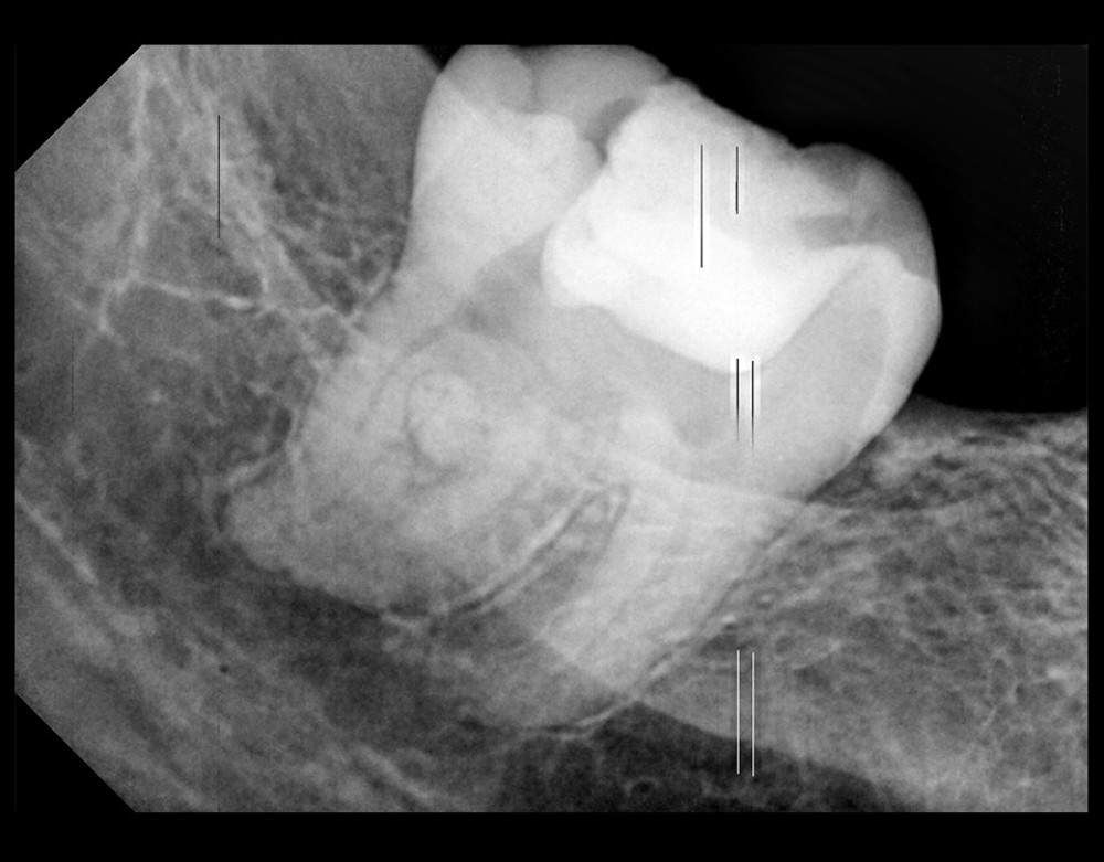
The man had a intraoral periapical radiograph of the lower right jaw performed which showed a fused wisdom tooth and supernumerary tooth with an irregular morphology and wide mesiodistal width. The x-ray made it hard to discern the demarcation between the pulp chamber of the wisdom tooh and the supernumerary tooth and the root canal configuration.

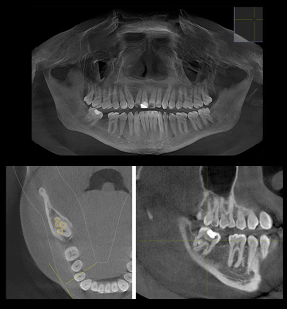
This led the man to have a cone-beam computed tomography (CBCT) imaging exam perormed to better view the tooth anatomy. The CBCT exam showed the pulp chamber was continuous and there were five canals in the fused tooth.

The man was presented with treatment options and the risks and benfits including extraction or root canal treatment. The man chose root canal treatment. After the root canal treatment was performed the tooth was restored with a temporary filling. A week later the man was to see his dentist to have a full coverage restoration performed. Six months later an x-ray was taken and showed normal apical tissue and there was no tenderness when touching. Further the follow-up exam did not show any swelling or sinus tract swelling.
In the discussion of the case the authors state that dental fusion may occur in up to 0.2% in permanent teeth. They state that cavity preparation when performing root canal treatment on a fused tooth can be a difficult procedure. While x-rays are helpful, they do not always show the complex anatomy needed for a successful procedure. The authors feel that CBCT provides a 3D view of the pulp chamber and evidence suggests that with complicated cases can provide significant additional information to the clinician which can ultimately modify the treatment plan and even diagnosis. Further a postion statement from the the American Association of Endodontists and the American Academy of Oral and Maxillofacial Radiology, suggest that CBCT should be considered the diagnostic modality of choice for dental anomalies.
The authors state:
“…we described the successful management of nonsurgical root canal treatment of fused third molars with supernumerary teeth that was performed using CBCT imaging. Successful treatment can be predicted when clinicians use a proper treatment plan and utilizing all available diagnostic tools.”
Currenty the origin of fused teeth remains unclear. However trauma in the area along with genetics are potential contributing factors
