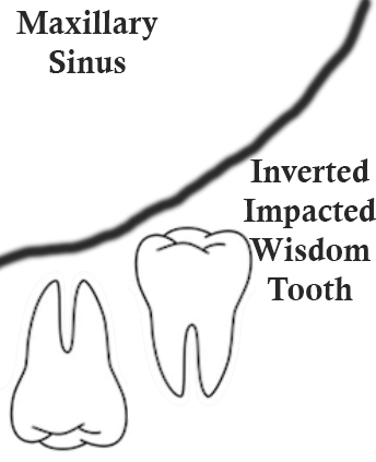
An interesting article titled “An unusual case of an inverted and impacted maxillary third molar,” written by Rammal and Alfonso appears in Oral Surgery in 2014 (vol. 7, pp. 109–111). The article discusses a case of an inverted and impacted upper wisdom tooth.
In the article the authors discuss how there are seven possible positions that a tooth can be classified into and one of these is the inverted position where the
the root apex faces the alveolar crest and the crown faces the maxillary sinus. In such a presentation the tooth appears as if it is growing into the maxillary sinus and the tooth clearly has no use for chewing in the mouth. The authors state:
While inverted impacted third molars [wisdom teeth] remain dormant without significant manifestation, removal presents difficulty because of the need to remove large amounts of bone and often present with the risk of displacement into the maxillary sinus.”
Displacement of a wisdom tooth into the maxillary sinus has been discussed as a possible complication of removal, see http://www.teethremoval.com/complications.html.
In the article the authors describe a 32 year old female who presented to the authors dental clinic complaining of pain in the upper right part of the mouth. She was found to have several cavities on teeth and a panoramic radiograph showed an inverted impacted upper left wisdom tooth. She did not report any pain in the upper left part of her mouth. She then had a cone beam computed tomography (CBCT) scan performed. The CBCT scan allowed for aiding in accurately diagnosing the position of the tooth in relation to anatomical structures such as the maxillary sinus and the zygomatic process of the maxilla. Furthermore the CBCT scan allowed for ruling out any pathology not seen on the panoramic radiograph.
The authors believed that if this inverted wisdom tooth was extracted complications were likely such as accidental displacement into the maxillary sinus, pterygomaxillary space, or the infratemporal fossa. This would then require another procedure to retrieve the displaced wisdom tooth. The authors seemed to suggest a conservative management approach of which the woman agreed since she did not have any symptoms. However, if symptoms appear then a more careful weighing of the pros and cons should be made.

Thus it would appear in cases of inverted impacted wisdom teeth and particularly inverted impacted upper wisdom teeth right next to the maxillary sinus retaining the wisdom tooth should be the preferred first option. The wisdom tooth should be evaluated with CBCT in addition to panoramic x-ray as the benefits of the additional diagnostic information provided outweigh the risks of the radiation exposure. CBCT can allow for accurately visualizing the exact location of the tooth in relation to anatomical structures to allow for surgical planning if removal becomes necessary. Removal should generally only be considered when symptoms warrant it. An additional procedure may then need to be undertaken to retrieve the wisdom tooth if it is displaced into any anatomical spaces. Of course the patient should fully be told of what is occurring and be able to weight the pros and cons of surgical extraction themselves.
