An interesting article titled “Traumatic myositis ossificans of the temporal
muscle after dental local anesthesia,” written by S.B. Helland and T.Ø. Pedersen appears in Clinical Case Reports (no. 11, pp. e7410, 2023). The article describes a case of a thirty year old woman who developed ossification of the temporal muscle attachment after local trauma during dental treatment which prevent her from opening her mouth.
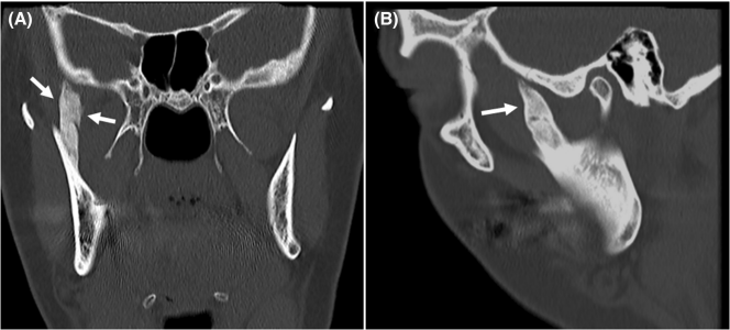
The thirty year old woman presented four months after having had a root canal treatment of a maxillary premolar tooth when local anesthesia was given near the right temporal muscle attachment. At the time she noted a sharp pain right after the injection was given. He also experienced persistent limited mouth opening after the dental treatment. Computer tomography was taken when the woman presented four months later which showed a bony growth roughly 3cm in length extending from the right coronoid process of the mandible to the cranial base.

Computer tomography in frontal (A) and sagittal (B) view showing ossification of the right temporal muscle attachment. From S.B. Helland and T.Ø. Pedersen, “Traumatic myositis ossificans of the temporal muscle after dental local anesthesia,” Clinical Case Reports, no. 11, pp. e7410, 2023, and has a Creative Commons license.
A coronectomy was performed to remove the bony lesion via an intraoral access. The woman was diagnosed with traumatic myositis ossificans. After surgery the woman was still not able to open her mouth as much as desired even though no bony interferences could be identified radiologically. The woman was given physical therapy and performed regular jaw exercise and five months later was able to achieve better mouth opening (27 mm). Five years later her mouth opening contined to improve and was back to the normal masticatory function range (40 mm).
The authors describe traumatic myositis ossificans (TMO) as very rare condition that can occur in the head–neck region. The masseter is the masticatory muscle most commonly affected, and most cases ocurring after surgery such as wisdom tooth removal. The onset of symptoms typically occurs 3 to 6 weeks after local muscle trauma. They say that the recommended treatment is to remove the ossified muscle to increase mouth opening. As the condition is rare, there is not a consensus of the preferred timing of surgery or if interpositional grafts should used.

Photo by Quang Tri NGUYEN on Unsplash
The authors suggest that dentists and dental patients consider traumatic myositis ossificans if patients experience resistant trismus after dental procedures. It is rare but some cases of TMO affecting the medial pterygoid muscle after nerve block, accomplished through local anestheisa, have been described.
