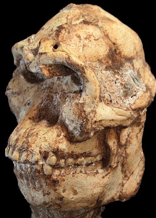
An interesting article titled “Preliminary paleohistological observations of the StW 573 (‘Little Foot’) skull” appears in eLife written by Amélie Beaudet and et al. (March 2, 2021). The article explores initial results an X-ray based imaging investigation of the skull of a 3.67-million-year-old Australopithecus specimen which is a prehuman ancestor known as Little Foot. In the study the authors presented initial results of the investigation of the dentition and cranial bones of Little Foot.
Little foot was found in a cave in the 1990s in South Africa. It took 15 years to completely remove Little foot from the ground and is regarded as the most complete skeleton of the early hominin lineage leading to humans. The researchers brought Little Foot’s skull from South Africa to the U.K. to scan it with a synchrotron x-ray microcomputed tomography system.
The authors were able to, for the first time, create a 3D spatial organization of the Haversian systems in the mandibular symphysis of an early hominin. They found that the dentine–cementum junction and the boundary between the cementum and the sediments were visible. The researchers found that lines and pits observable on the distal-buccal aspect of reconstruction were located at about 2.4 and 3.6 mm from the cemento-enamel junction, and using the synchrotron-based data sets the color map showed that they were slightly less clearly rendered in the 3D reconstruction. This indcates that reductions in the normal thickness of enamel correspond to disruptions to the normal growth of enamel and indicate two disruptive events in the life history. These life events could have been periods of dietary deficiency, nutritional stress, or illness.

The authors also observed less than 1-mm channels in the braincase that may have been used for brain thermoregulation. Modern human brains are about three times bigger than Little Foots was so the information the researchers determine could help to answer how brain temperature regulation has evolved during that time. The researchers also were able to observe the vascular canals in the mandible, which may uncover the biomechanics of how Little Foot ate food. The authors were very encouranged by the imaging results that they obtained using synchrotron radiation.
The authors plan to continue with their imaging process of Little Foot. They are hopeful that it can help to answer questions regarding compact bone microstructures to help provide insights into the evolution of the bone modeling/remodeling process.
